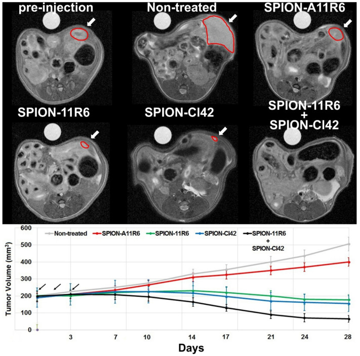
Figure 5.
In vivo aptamer-conjugated SPIO nanoparticle therapeutic efficacy assessed using noninvasive MRI. Representative axial MR images (upper row) and corresponding quantitative measurements of tumor volume (lower row) in 4T1 tumor-bearing mice. Noninvasive imaging protocols were performed pre- (t = 0; corresponding to three-weeks post-tumor cell inoculation in the mammary fat pad) and up to 28 days post-injection of three consecutive doses (24 h interval time) of different aptamer therapeutic formulations. Black arrows highlight the injection time points. Pre-injection, non-treated mice, SPION-A11R6, SPION-11R6, SPION-Cl42RNA, and combined SPION-11R6 + SPION-Cl42RNA mice injected groups. White arrows reveal the tumor location and size highlighted with red contours. Data are expressed as mean ± SD, n = 3 per group.

