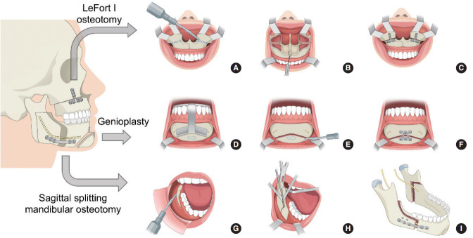
Fig. 2.
Schematic drawing of maxillomandibular advancement. LeFort I osteotomy. (A) Exposure of the piriform aperture and nasal floor. Le Fort I osteotomy and wedges (dotted lines) for counterclockwise rotation. (B) Maxilla mobilization via traction with wire through the anterior nasal spine. (C) Fixation with titanium plates. Virtual surgical planning-guided genioglossus and geniohyoid advancement. (D) The anterior mandible is exposed and the osteotomy guide is designed to capture the genial tubercle (dotted line) while avoiding dental roots and mental nerves. (E) The osteotomy is made using a reciprocating saw. The genial tubercles (dotted line) are preserved. (F) The mobilized graft is moved forward and fixed with a plate. Sagittal split mandibular osteotomy. (G) The mandibular ramus and body are exposed by subperiosteal dissection. (H) The horizontal osteotomy is made through the lingula and the anterior osteotomy split sequentially with three osteotomes. (I) The mandible is advanced, and fixation is performed with positional screws and titanium plates.

