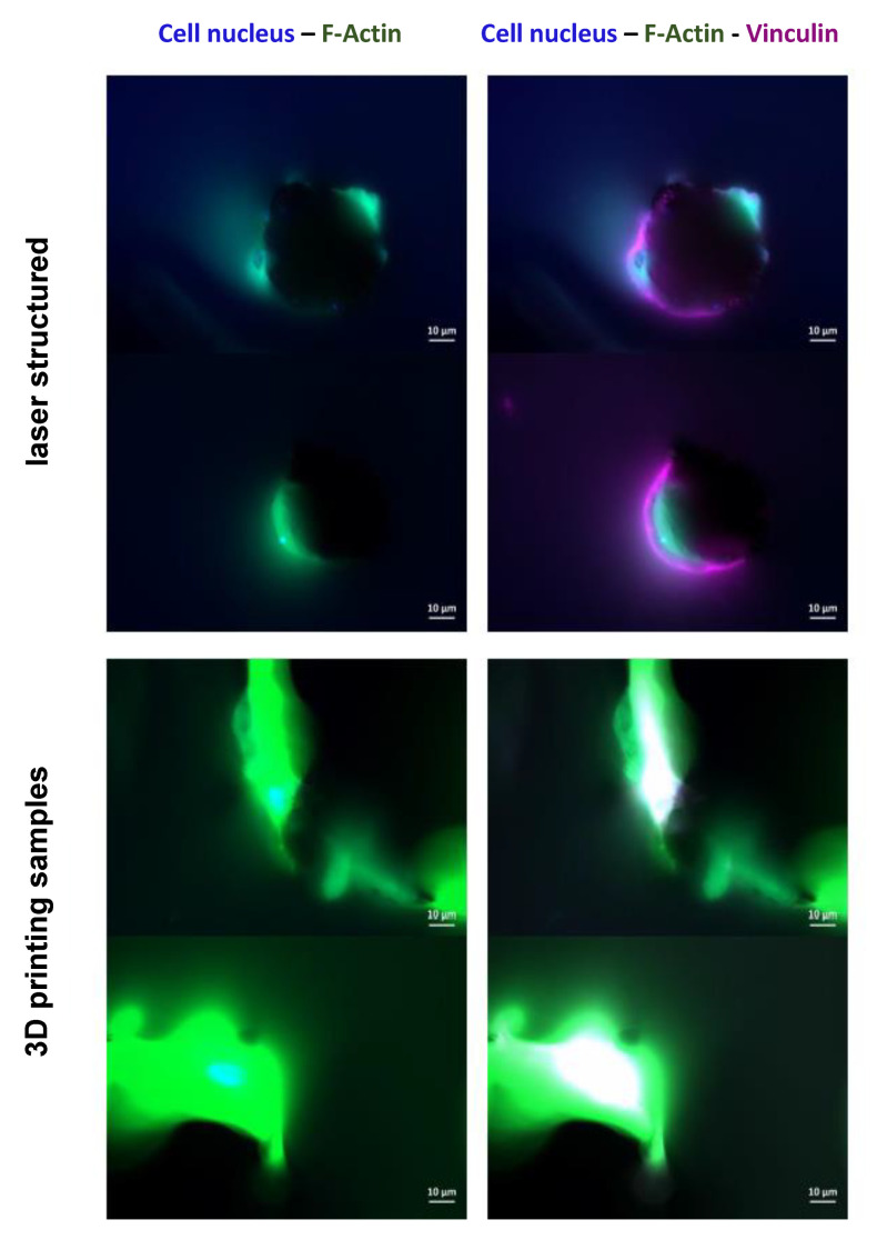
Figure 4.
Immunofluorescence staining of human bone marrow stroma cells (hBMSCs) 24 h after sowing. Cell nuclei appear in blue (DAPI staining). Due to the three-dimensionality of the surfaces, the images lack clear sharpness and provide only a representation of cellular structures. Fibrillary actin (F-actin; green fluorescence)- and vinculin (violet fluorescence)-positive cells are shown.

