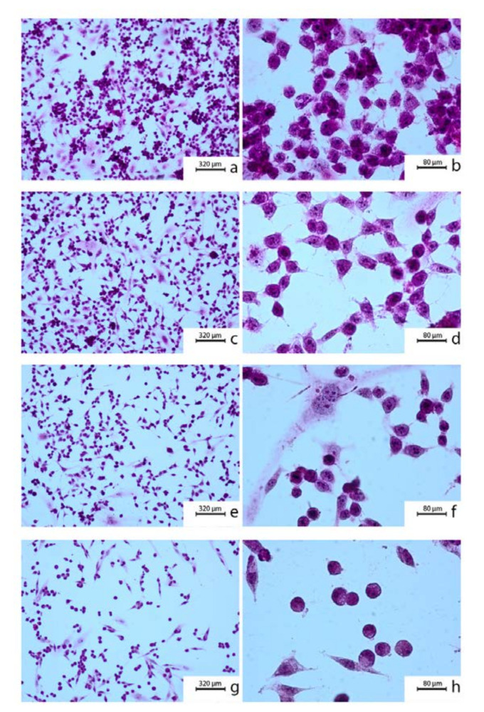
Figure 1.
Cytomorphological view of OV7 cells after 24 h incubation: (a,b) control (no treatment); (c,d) 10 μM caffeic acid phenethyl ester (CAPE); (e,f) 50 μM CAPE; (g,h) 100 μM CAPE. To prepare the samples a hematoxylin and eosin staining was used. Exposition: optical magnification ×100 (a,c,e,g), ×400 (b,d,f,h). Main features: cellular polymorphism, hyperchromasia, long cytoplasmic protrusions (a,b); irregular nuclear shapes, numerous nucleoli visible, strong cellular adhesion (c,d); densification of the nuclei; shortening of cytoplasmatic protrusions (e,f); round cells, poorly attached to the surface (g,h).

