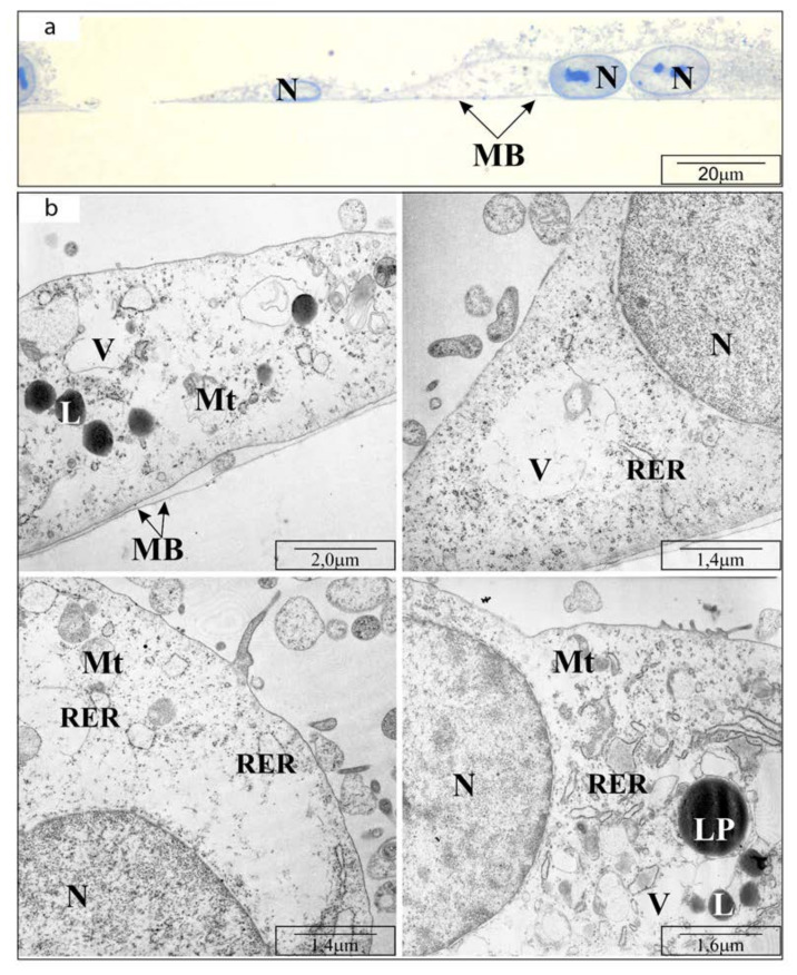
Figure 3.
Transmission electron microscope visualization of OV7 cells treated with 25 µM CAPE, after 24 h of incubation: (a) cross-section through the monolayer of cultured cells (half-thin sections, i.e., 0.5 μm thick); (b) cell organelles: MB—basement membrane; N—cell nucleus; RER—rough endoplasmic reticulum; L—lysosomes; LP—lipoprotein bodies; Mt—mitochondria (highly deformed); V—vacuoles. Short description: cell enlargement, decrease in osmophilicity.

