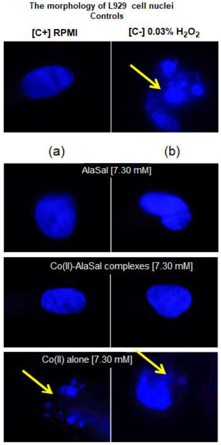
Figure 7.
Microscopic images of L929 cells with a sign of cell nuclei damage. Cell cultures in complete RMPI-1640 medium (cRPMI) were used as positive control (C+): cells with no sign of cell nuclei damage; cells treated with 0.03% H2O2 were used as negative control (C−): cells with DNA damage. L929 cells stimulated with selected compounds: (a) solution prepared freshly or (b) solution stored for two weeks. The morphology of cell nuclei was assessed by 4′,6-diamidino-2-phenylindole (DAPI) staining. Samples were viewed under a fluorescent microscope (Axio Scope A1, Zeiss).

