Figure 1.
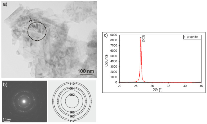
Transmission electron microscopy (TEM) micrograph (a) and selected area electron diffraction (SAED) pattern of the area marked as A in Figure 1a with identification (b), as well as the X-ray diffraction (XRD) pattern (c) of graphite particles consisting of graphene layers stacked together.

