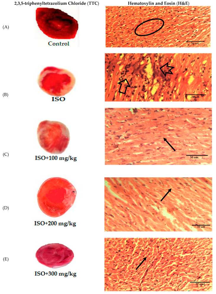
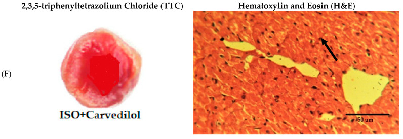
Figure 13.
Effect of Anogeissus acuminata Cr on representative photomicrograph of heart tissues dyed with 2,3,5-triphenyltetrazolium chloride (TTC) (left) and Hematoxylin and Eosin (H&E) stain (right) in ISO-induced myocardial infarction at (A) control, (B) intoxicated ISO, (C) 100 mg/kg, (D) 200 mg/kg, (E) 300 mg/kg and (F) standard (Carvedilol) in Sprague Dawley rats. White color in TTC staining represents area of necrosis, while in H&E staining, oval circle represents normal cells, arrow shows edema, notched arrow depicts necrosis with lost nuclei and simple arrow shows dense normal cluster of cardiac myocytes (n = 6).


