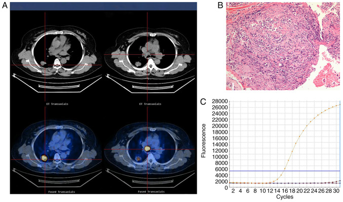
Figure 1.
Representative images of an adenocarcinoma with wild-type EGFR in a 55-year-old female who had never smoked with normal serum CEA levels (3.5 ng/ml). (A) 18F-FDG PET/CT in the axial plane and whole body maximum-intensity projection images, demonstrating abnormal FDG uptake in a left upper lobe tumor (SUVmax, 17.4) and the SUVmax of the mediastinal lymph node, 21.7. (B) Hematoxylin and eosin-stained tissue indicating adenocarcinoma features. Magnification, ×100. (C) Polymerase chain reaction confirmation of the EGFR wild-type. EGFR, epidermal growth factor receptor; SUVmax, maximum standardized uptake value; CEA, carcinoembryonic antigen; 18F-FDG PET/CT, 18F-fluorodeoxyglucose positron emission tomography-computed tomography.

