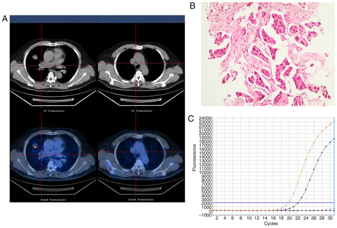
Figure 2.
Representative images of an adenocarcinoma with EGFR exon 19 deletion in a 61-year-old male who had never smoked with abnormal serum CEA levels (9.8 ng/ml). (A) 18F-FDG PET/CT in the axial plane and whole body maximum-intensity projection images, demonstrating abnormal FDG uptake in a right upper lobe tumor (SUVmax, 10.6) and the SUVmax of mediastinal lymph node was 4.5. (B) Hematoxylin and eosin-stained tissue indicating adenocarcinoma features. Magnification, ×100. (C) Polymerase chain reaction confirming the EGFR exon 19 deletion. EGFR, epidermal growth factor receptor; SUVmax, maximum standardized uptake value; CEA, carcinoembryonic antigen; 18F-FDG PET/CT, 18F-fluorodeoxyglucose positron emission tomography-computed tomography.

