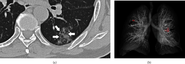
Figure 8.

A 76-year-old man presented without onset symptoms. Axial CT image shows patchy ground-glass opacities located at the lateral basal segment of the left lower lobe (a). The interlobular septa (white arrow) are thickened and show a reticular pattern. The 3D reconstruction image shows bilateral opacities (red spot and black arrow) with peripheral distribution (b).
