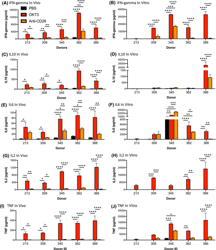
FIGURE 3.

Comparison of cytokine release using the in vivo PBMC‐NSG model and an in vitro PBMC assay. PBMC from five donors were evaluated for in vivo (A, C, E, G, and I) and in vitro (B, D, F, H, and J) cytokine release. For in vivo cytokine release, irradiated (100 cGy) NSG mice were injected IV with PBMC (20 × 106 cells) and 6 days post‐injection, mice were treated IV with PBS, OKT3 (0.5 mg/mL), or anti‐CD28 (1 mg/mL) as described in the Materials and Methods. Six hours after treatment sera were collected for human cytokine analysis. For in vitro cytokine release, PBMC were added to plates that were coated overnight with PBS, OKT3, or anti‐CD28 as described in the Materials and Methods. After 48 hours supernatants were collected for human cytokine analysis. Cytokine quantification included: (A, B) IFN‐gamma; (C, D) IL10; (E, F) IL6; (G, H) IL2; (I, J) TNF. Cytokine levels (pg/mL) ± SEM are shown. Each in vivo experimental group represents three to five mice, and in vitro results were performed in duplicate for each donor. *P < .05, **P < .01, ***P < .001, ****P < .0001
