
Figure 1.
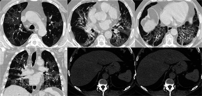
Chest CT severity score and CTL/S. Non‐enhanced CT images in a 57 years‐old male with COVID‐19 pneumonia. Axial images in upper, mid and basal portions of the thorax (figs. a‐c) and coronal reformation (fig. d) demonstrate multiple and bilateral ground‐glass opacities that involve more than 50% of some segments. These infiltrates are typical of COVID‐19 infection, categorized as CO‐RADS 5. The chest CT severity score was 36. The upper abdominal axial images from the same data set (figs. e‐f) showed decrease liver attenuation (ROIs average of 29 UH) when compare to spleen (45 HU). The CTL/S ratio was 0.65