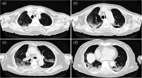
Figure 1.

Representative images of thoracic computed tomography (CT) reveal ground‐glass opacity, which shows segmental distribution, (a) predominant in the subpleural area of the lateral‐dorsal side (b and c) of the lower lobe (c and d). The right lobe seems predominantly affected (a–d).
