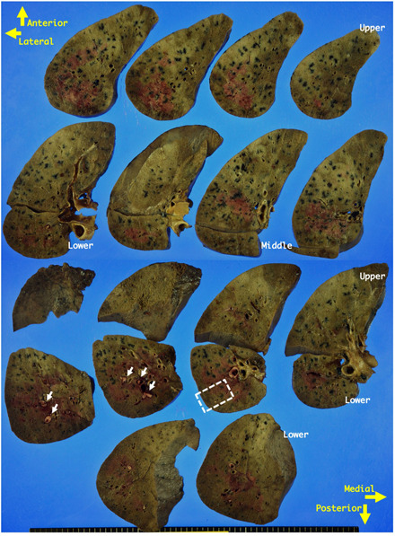
Figure 3.

Gross appearance of transverse sections from the right lung. The cut surfaces appear glittering and focally brownish‐red and are solid and firm. Adjacent to the brownish‐red areas, yellowish‐white thrombi are seen in interlobular vessels (arrows). The changes are unevenly distributed and appear fuzzy segmentally. The subpleural area on the lateral‐dorsal side appears more solid and firm. The area distant from the pleura is soft and has much more exudate. The area marked with a square is representative and has been used to present the histological appearances in Fig. 4.
