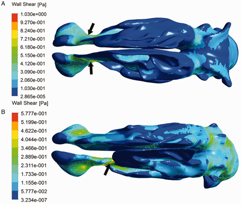
Figure 3.
The contour of wall shear stress. (A) Patient of NSD with the formation of CB, the higher value was located near the nasal valve area (arrows) and no evident difference of wall shear stress distribution between bilateral nostril sides. (B) Patient of NSD without CB, the overall wall shear stress in deviated side was higher than that in nondeviated side, and the location of maximal wall shear stress in deviated side was also transferred (arrow).

