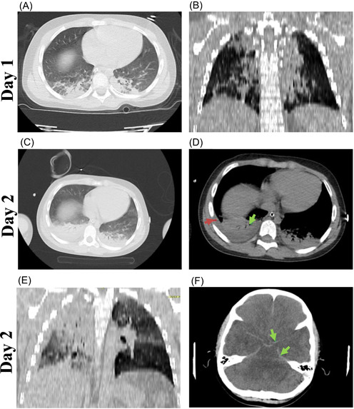
Figure 1.

A 9‐year‐old boy presented with cardiopulmonary arrest, fever, nausea, abdominal pain, headache, anorexia, and fatigue. Day 1: A, Axial computed tomographic (CT) scan in parenchymal view showing consolidation at both posterior basal segments with air bronchogram sign. B, Coronal reconstructed CT image in parenchymal view showing consolidation in both lungs. Day 2: C, Axial CT scan in parenchymal view showing consolidation at both posterior basal segments with air bronchogram sign, which has progressed, in comparison to day 1. D, Axial CT scan in mediastinal view showing consolidation with air bronchogram sign (green arrow). Also, a mild pleural effusion at the right side (red arrow). E, Coronal reconstructed CT image in parenchymal view showing consolidation in both lungs, more prominent at right hemithorax. F, Axial CT scan of the brain showing hyperdensity at basal cisterns, interhemispheric, and bilateral Sylvian fissures in favor of subarachnoid hemorrhage, without intraventricular hemorrhage and hydrocephalus (green arrow); decreased density of white matter in favor of brain edema
