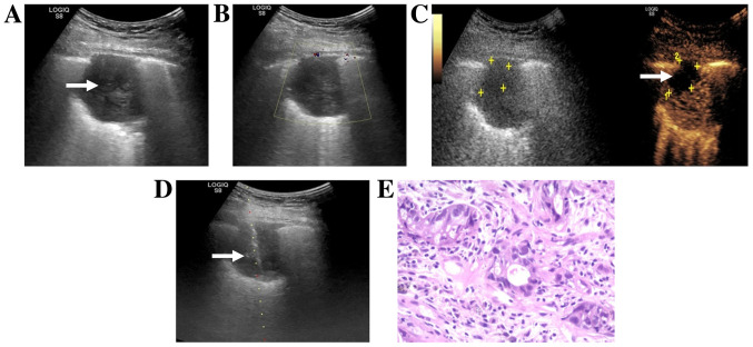
Figure 3.
Images from a 47-year-old man with a history of chest pain and hemoptysis. (A) Routine ultrasound showed a hypoechoic lesion (white arrow) in the superior lobe of the right lung. (B) Color Doppler flow imaging showed no color flow signal around or inside the nodule. (C) CEUS obtained 42 sec after an injection of SonoVue® (Braggo) showed irregular necrosis in the anterior part of the lesion (white arrow and yellow markings). (D) US-guided transthoracic biopsy passed through the necrotic area and targeted the enhanced area (white arrow). (E) Hematoxylin-eosin staining of histopathological biopsy specimen revealed an adenosquamous carcinoma (original magnification, ×200). The H&E staining results were obtained using routine histopathology. CEUS, contrast-enhanced ultrasound; US, ultrasound.

