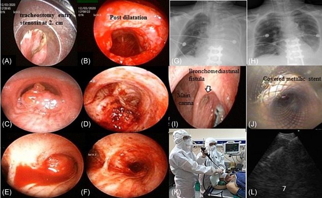
Figure 2.

Examples of cases in the study, A‐J. (A and B: pre and postprocedure view of postintubation tracheal stenosis; C and D, pre and postprocedure view of tracheal malign airway obstruction with adenoid cystic carcinoma; E and F, pre and postprocedure view of central (right main bronchus) malign airway obstruction with squamous cell carcinoma; G and H, pre and postprocedure chest X‐ray of left lung complete atelectasis with mucus plaque; I and J, bronchomediastinal fistula, and after covered with metallic stent). K, Protection of health workers and patient's during EBUS‐TBNA L: sonographic view of a sampled subcarinal lymph node. EBUS‐TBNA, endobronchial ultrasonography guided‐transbronchial needle aspiration [Color figure can be viewed at wileyonlinelibrary.com]
