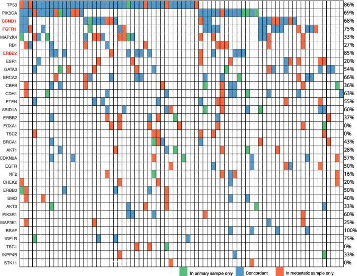
Fig. 3.
Concordance plot of primary and metastatic paired samples. Data for 76 patients and their paired sequenced samples is shown. Genetic alterations included in this figure are putative driver genes observed in 2 or more samples. A further 4 patients and their paired samples have been excluded from the figure because their pairs did not include the genes shown. Concordance scores for each gene is listed on the right of the figure. Genes listed in red indicate that it is CN amplification concordance being compared, rather than mutation concordance (shown in black text)

