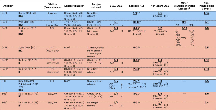
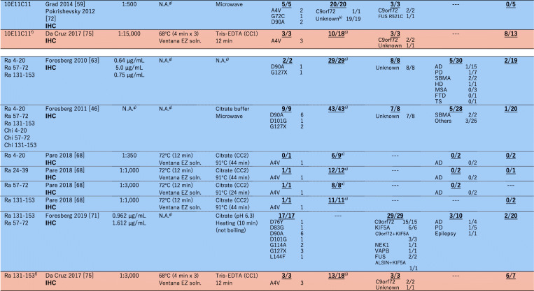
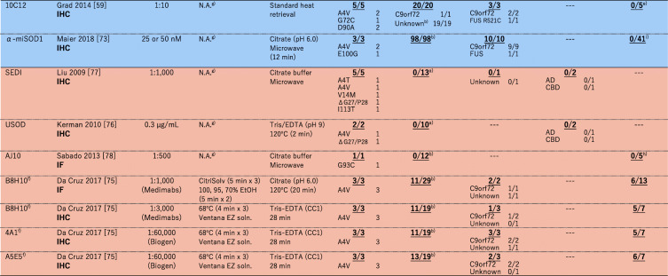
Table 1.
A summary on immunohistochemical (IHC)/immunofluorescence (IF) detection of misfolded SOD1 with misfolded-SOD1 antibodies in human spinal cords. The numbers in each box represent (the number of misfolded-SOD1-positive cases)/(the number of total cases examined)
aNo SOD1 mutations were confirmed. bThere is no mention on the presence or absence of SOD1 mutations. cNo motor neurons in the five remaining cases. dAD, Alzheimer’s disease, PD Parkinson’s disease, DLB Dementia with Lewy body, PSP Progressive supranuclear palsy, FTD Frontotemporal dementia, NIFID neuronal intermediate filament inclusion disease, SBMA Spinal and bulbar muscular atrophy, HD Huntington’s disease, MSA Multiple system atrophy, TS Tuberous sclerosis, CBD Corticobasal degeneration. eThere is no mention on the non-neurological controls in the paper. fIn this review, the cases with cytoplasmic granular staining, rare round deposits, abundant round deposits, and globular inclusions are counted as misfolded-SOD1 positive, while the cases with no signal, sparse diffuse staining, and abundant diffuse staining are counted as misfoded-SOD1 negative. gNot available (no mention in the paper). hThe control cases are described as “non-ALS controls”. iIn the paper, it was described that “no or only weak immunoreactivity was observed in motor neurons of most of the 41 spinal cord tissue samples from NNC patients”


