Fig. 1.
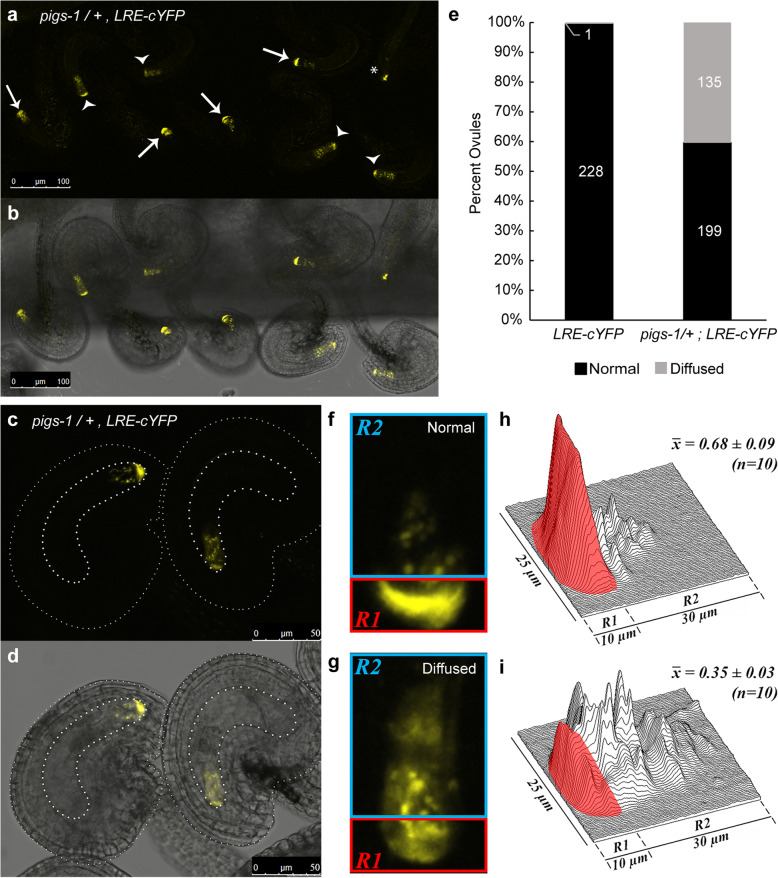
pigs-1 mutation disrupts polar localization of LRE-cYFP in the filiform apparatus of synergid cells. a-b Localization of the LRE-cYFP fusion protein in a pigs-1/+ pistil. a Fluorescent image of a portion of the pistil captured in the YFP channel of a confocal microscope and b Merged view of fluorescent image in (a) with the bright field image of the same portion of the pistil. White arrows, FGs with a polarized cYFP localization in the filiform apparatus; white arrowheads, sibling FGs with a diffuse cYFP localization throughout the synergids; Asterisk, FG not scored for localization due to obscured view from mispostioning of the ovule during mounting on a slide. Bar = 100 μm. c-d Enlarged view of two ovules within a pigs-1/+ pistil Bar = 50 μm. c Fluorescent image captured in the YFP channel of a confocal microscope and d Merged view of fluorescent image in (c) with the bright field image of the same ovules captured in (c). e Percent ovules showing normal and diffuse LRE-cYFP localization in synergid cells of wild-type and pigs-1/+ pistils. Numbers in the column refer to total number of ovules scored for indicated categories. f-g Representative images of (f) normal and (g) diffuse LRE-cYFP localization patterns scored within synergid cells of a pigs-1/+ pistil. h-i Surface plots of (h) normal and i diffuse LRE-cYFP fluorrescent signal scored within synergid cells of a pigs-1/+ pistil. X̅ values are average raw integrated densities of cYFP signal in filiform apparatus portion of the synergid cell boxed in red (R1) divided by total signal in R1 + R2, where R2 represents remainder of the synergid cell boxed in blue

