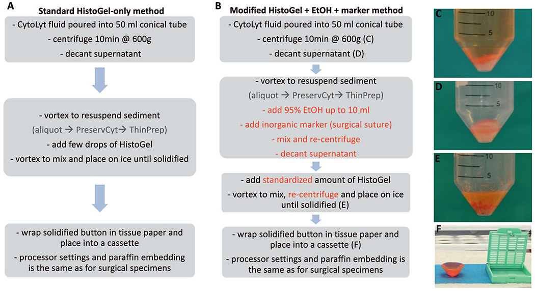
Figure 1.

Protocol flow charts for the standard HistoGel method (A) versus the modified HistoGel + ethanol (EtOH) method (B). Key steps in each protocol are outlined. The modified steps are in a red font. The photographs illustrate initial sediment before (C) and after (D) decanting of the supernatant, sediment mixed with HistoGel after solidification on ice (E), and the typical appearance of the resultant solidified button before placement into a cassette (F).
