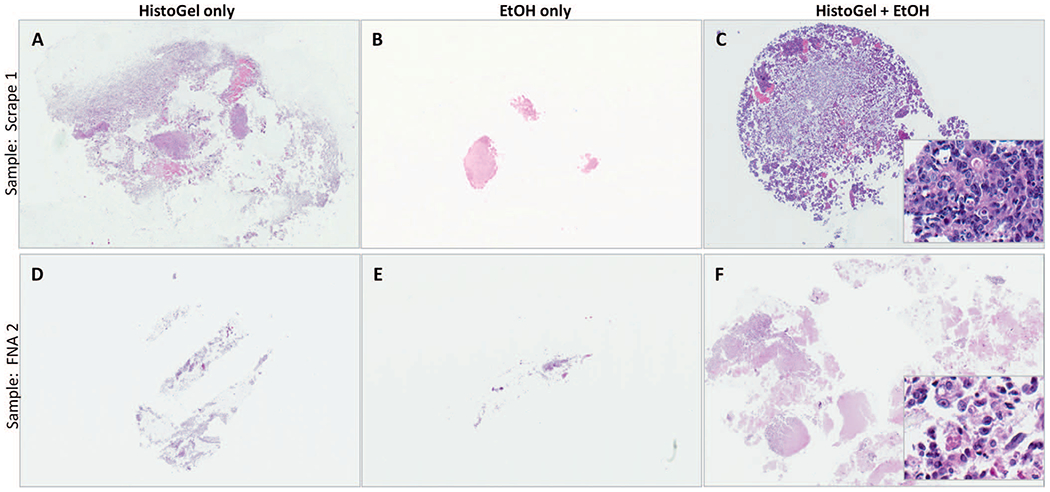
Figure 3.

Illustration of the microscopic appearance of cell blocks prepared by the HistoGel + ethanol (EtOH) method versus HistoGel-only and the EtOH-only methods. Note the substantially greater cell capture and more-compact appearance of cell blocks prepared with the HistoGel + EtOH method. For quantitation of increased cell capture, see Table 1. The insets in C and F illustrate excellent cytomorphologic preservation with this method (hematoxylin-eosin, original magnifications ×20 [A through F] and ×400 [insets]).
