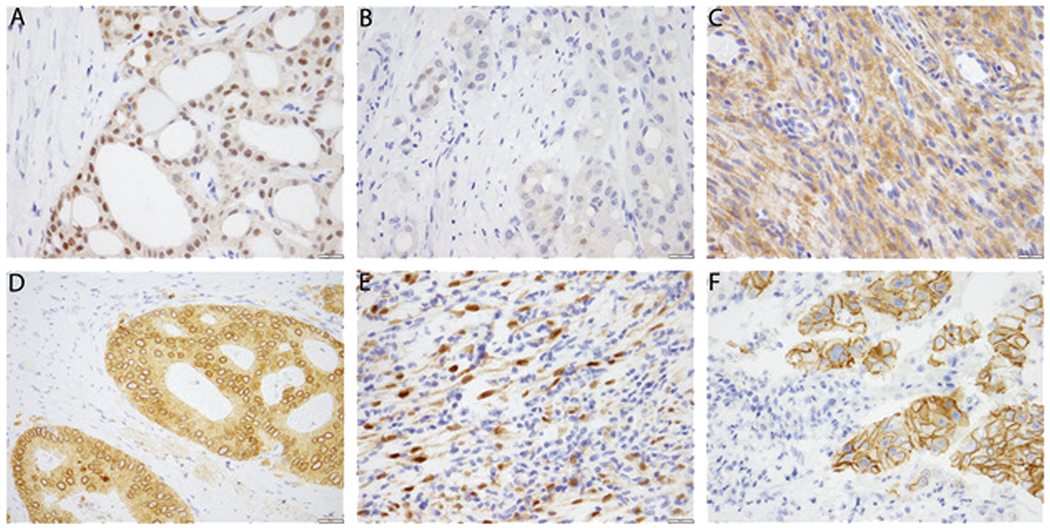
Fig. 1.

Patterns of pan-Trk immunohistochemistry expression in NTRK fusion positive cancers. a Strong cytoplasmic and nuclear staining in this secretory carcinoma of the salivary gland with canonical ETV6-NTRK3 fusion. b Staining is occasionally weak and focal, seen in approximately 1% of tumor cells, as in this secretory carcinoma of the salivary gland. c Cytoplasmic staining is seen in this Lipofibromatosis-like Neural Tumor with a TPM3-NTRK1 fusion. d Cytoplasmic and perinuclear staining is seen in this colonic adenocarcinoma with a LMNA-NTRK1 fusion. e Cytoplasmic and nuclear staining is seen in this inflammatory myofibroblastic tumor with an ETV6-NTRK3 fusion. f Membranous staining is seen in this intrahepatic cholangiocarcinoma with a PLEKHA6-NTRK1 fusion
