
Fig. 2.
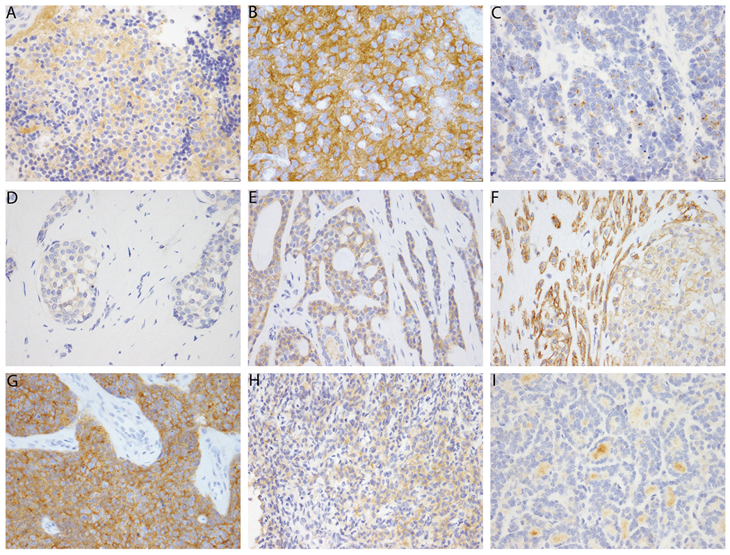
Pan-Trk immunohistochemistry expression in NTRK wild type carcinomas. a, b This pitfall can be seen in neural derived tumors such as neuroblastoma (a) and oligodendroglioma (b). c Occasionally focal cytoplasmic staining can be seen in carcinoma with neuroendocrine differentiation. d Weak cytoplasmic staining is seen in a minority of breast invasive ductal carcinomas. e Adenoid cystic carcinoma often shows moderate to strong cytoplasmic staining. f Cytoplasmic and membranous staining is seen in this atypical pleomorphic adenoma. The myoepithelial cells show particularly strong staining. g, h There is often staining in sarcomas, particularly those with neural or smooth muscle differentiation or those with other translocations, such as this desmoplastic small round cell tumor (g) and this sarcoma with BCOR-CCNB3 translocation (h). i In this papillary thyroid carcinoma, there is nonspecific staining of the colloid, but the tumor cells themselves show no staining, and the stain is therefore interpreted as negative