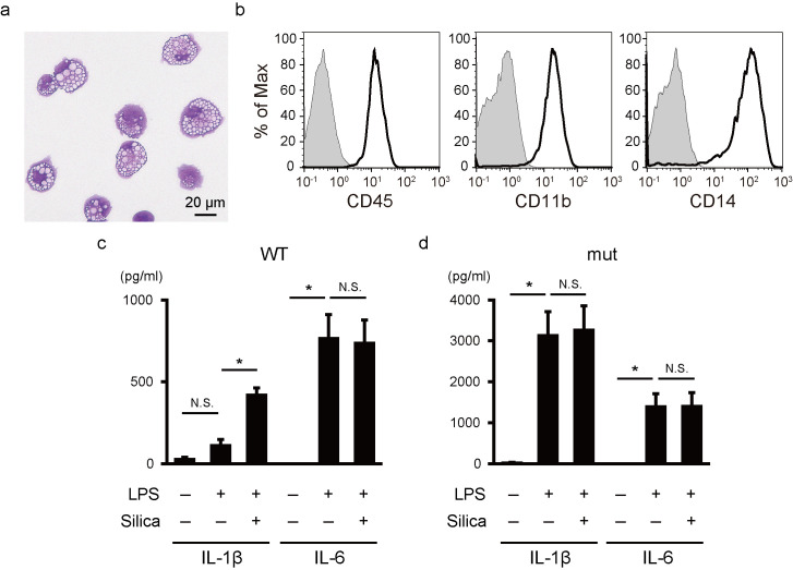
Fig 1. Functional monocytic cells on feeder- and serum-free monolayer culture.
(a) A representative May-Giemsa staining image of monocytic cells derived from iPSCs. (b) A flow cytometric analysis of monocytic cells. The staining profiles of specific antibodies (thick lines) and isotype-matched controls (gray areas) are shown. (c,d) IL-1β and IL-6 secretion from monocytic cells with wild-type (c) and mutant (d) NLRP3. Cells were stimulated with LPS (20 ng/ml) for 6 hours and silica (500 μg/ml) for an additional hour. Bars show the mean ± S.E.M. of four experiments. * P < 0.05 (paired t-test).

