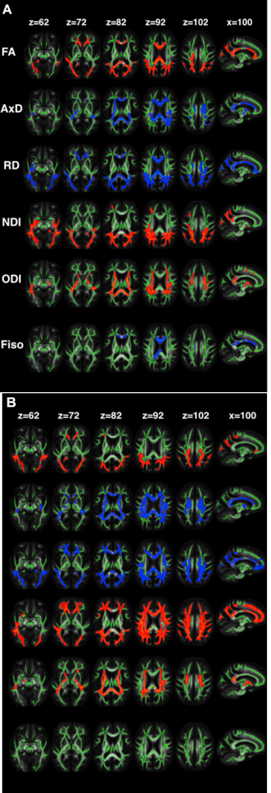
Figure 3.

The changes in diffusion tensor imaging (FA, axial diffusivity, and RD), neurite orientation dispersion, and density imaging (NDI, ODI, and Fiso) metrics in patients with (A) ε4– and (B) ε4+ young-onset Alzheimer’s disease in comparison with controls.
In the voxel-wise group differences, the metrics that are increased and decreased in patients are highlighted in blue and red, respectively. The results are overlaid on axial and sagittal planes of the white matter skeleton (shown in green) in the neurologic convention. Reproduced with permission from Slattery et al., 2017 (CC BY 4.0). AxD: Axial diffusivity; FA: fractional anisotropy; Fiso: fraction of isotropic water; L: left; NDI: neurite density index; ODI: orientation dispersion index; R: right; RD: radial diffusivity.
