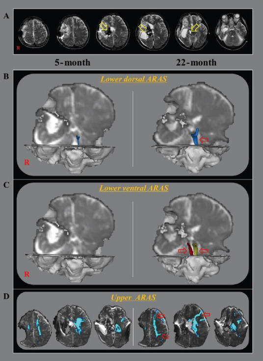
Figure 1.

DTT for the ARAS of a 33-year-old male stroke patient.
(A) DTT images taken at 5 months after onset reveal leukomalactic lesions in the right basal ganglia and thalamus, and midbrain (yellow arrows). (B) On 5-month and 22-month DTT images, the right lower dorsal ARAS was not reconstructed on both hemispheres, while thin left lower dorsal ARAS observed on the 5-month DTT image had become thicker on the 22-month DTT image (red arrow). (C) On 5-month DTT image, the lower ventral ARAS was not reconstructed in the both hemispheres, however, it was well-reconstructed on both sides on 22-month DTT image (red arrows). (D) On 5-month DTT image, the neural connectivity of the upper ARAS between the ILN and cerebral cortex was decreased in bilateral prefrontal cortex, parietal cortex, bilateral basal forebrain, and the right thalamus. By contrast, the neural connectivity of the upper ARAS was increased in the left prefrontal cortex and parietal cortex on the 22-month DTT image (red arrows). ARAS: Ascending reticular activating system; DTT: diffusion tensor tractography; ILN: intralaminar nucleus; R: right.
