
Figure 2.
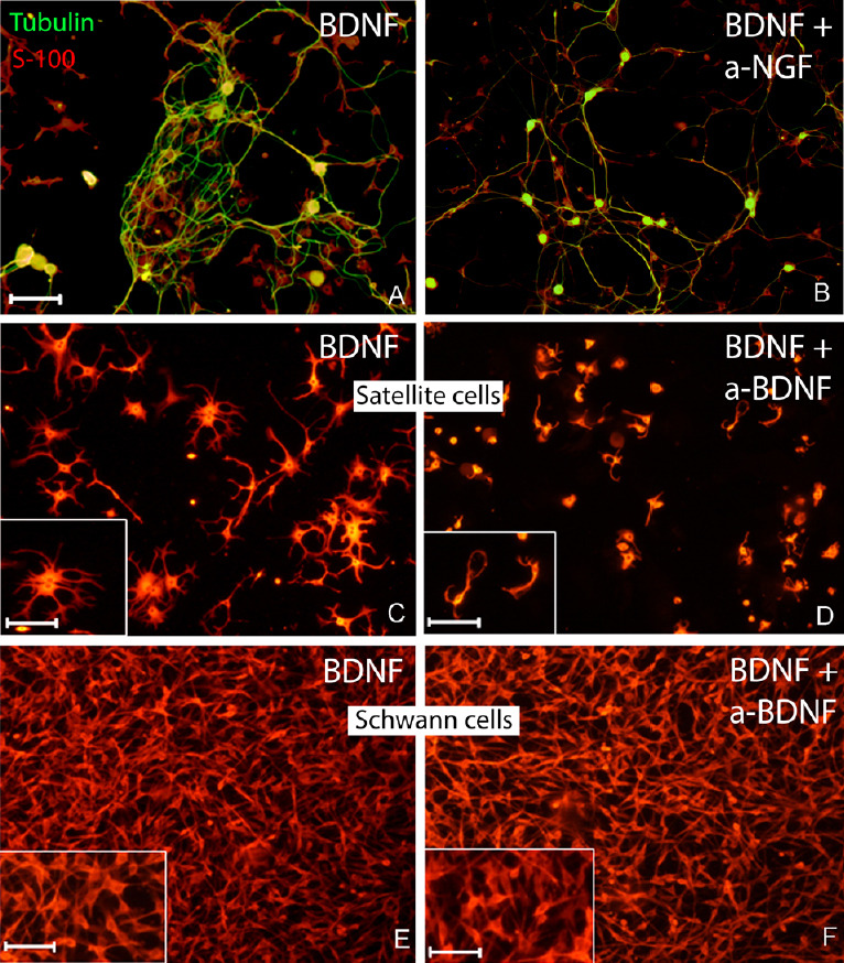
BDNF enhances cell cluster formation via activation of the satellite cells.
(A) In the presence of BDNF at 72 hours, a semi-defined cluster of β-tubulin-positive neurons (green) and S-100-positive satellite cells (red) was seen. (B) The cell cluster formation was avoided when BDNF and anti-nerve growth factor antibody were simultaneously added to the medium. BDNF specifically activated satellite cells and did not affect Schwann cells. (C, D) At 72 hours, S-100-positive satellite cells derived from dissociated DRG featured larger cell bodies and well-developed processes (C) as compared to the same culture with anti-BDNF antibody added to the medium (D). Both insets in C and D are higher magnifications to appreciate the more extensive cellular processes branching in the presence of BDNF compared to BDNF + anti-BDNF antibody. The immunostaining using S100 antibody did not reveal any morphological differences (in number, size, maturation features) in sciatic nerve-derived Schwann cell population cultured in the presence of BDNF (E) or without BDNF (F). Scale bars: 100 µm for A, F; 50 µm for all insets. BDNF: Brain-derived neurotrophic factor.