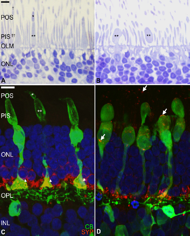
Figure 3.

Human photoreceptor degeneration process in an organotypic culture of the neuroretina.
Organ retinal explant cultures are considered useful tools for cellular and molecular research into retinal degeneration and neuroprotection. Briefly, human neuroretina explants were cultured in Transwell® plates, with the photoreceptor layer facing the supporting membrane. Ultrathin and cryostat sections were evaluated after toluidine blue staining (A, B) and after immunostaining for neuronal markers (C, D). Fresh human neuroretina (A) morphologic organization of the photoreceptors show easily recognizable cone and rod outer (asterisk and dagger, respectively) and inner segments (double asterisk and double dagger, respectively), outer limiting membrane, and highly organized outer nuclear layer. After 6 days of culture (B), the photoreceptor degeneration process is evident with loss of the cone outer segments and swollen cone inner segments (double asterisk) and cell bodies. Immunostaining for calbindin (CB, green), a calcium-binding protein of cones and second-order neurons (C), shows the normal morphology of the cone photoreceptors, including the outer (asterisk) and inner (double asterisk) segments and their terminals (arrowheads). After 9 days of culture (D) some inner segments are swollen, and the cones have degenerated inner and outer segments. Synaptophysin (SYP, red), a synaptic-vesicle protein in the photoreceptors and second-order neurons, was located at photoreceptor axon terminals (C) and after culture, it is identified throughout the photoreceptor cell bodies (D, arrows). Scale bars: 10 µm. These images were obtained in collaboration with Dr. Nicolas Cuenca (Universidad de Alicante, Spain). INL: Inner nuclear layer; OLM: outer limiting membrane; ONL: outer nuclear layer; OPL: outer plexiform layer; PIS: photoreceptor inner segment; POS: photoreceptor outer segment.
