
Fig. 5.
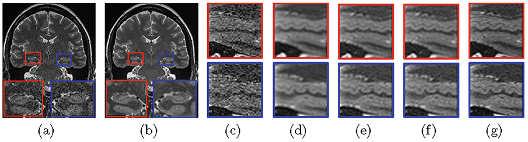
Reconstructed results on the OSC data set. Two coronal slices from (a) an LR image and (b) our reconstruction. The hippocampus in sagittal slices from (c) an LR image, and the images reconstructed via (d) Tikhonov, (e) TV, (f) MSTV, and (g) the proposed approach, respectively.