Figure 3.

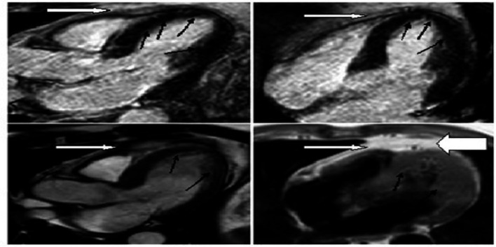
Cardiac MRI of the patient with perfusion showing normal left ventricle muscle (black arrows). The pericardium, pointed by white arrows, shows mild thickening (bold white) with no effusion.

Cardiac MRI of the patient with perfusion showing normal left ventricle muscle (black arrows). The pericardium, pointed by white arrows, shows mild thickening (bold white) with no effusion.