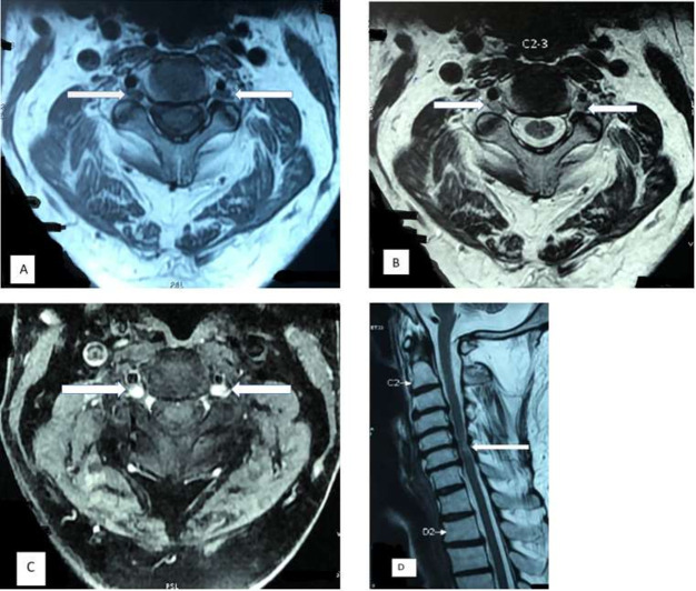
Figure 1.
MRI of the cervical spine in axial plane showing T1-weighted hypointense (A) and T2-weighted hyperintense (B) with T1-weighted Short Tau Inversion Recovery (STIR) post contrast enhancing (C) lesions at the C2–C3 vertebral level suggestive of dorsal root ganglionitis (arrows). (D) MRI in sagittal plane showing the T2-weighted hyperintense lesion in the spinal cord at the level of C6 vertebra suggestive of myelitis (arrow).

