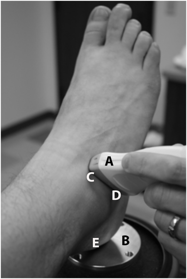
Fig 1. Sound head placement on the lateral STJ.
“A = Ultrasound probe; B = Vibration generator platform; C = End of ultrasound probe positioned over the lateral talar neck; D = End of ultrasound probe positioned over the distal lateral calcaneus; E = Calcaneal point of contact over the vibration generator”.

