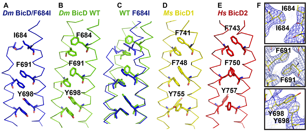
Figure 3. Conformation of key aromatic residues in Dm BicD-CTD/F684I.

(A) The Cα-trace of the structure of Dm BicD-CTD/F684I is shown (blue). Residues I684, F691, Y698 are labeled and shown in stick representation. (B) Structure of the wild-type Dm BicD-CTD (green, PDB ID 4BL6).9 (C) Least squares superimposed structures of Dm BicD-CTD/F684I and the wild type. (D, E) Structures of the Dm BicD homologs (D) Ms BicD1-CTD (yellow, PDB ID 4YTD)10 and (E) Hs BicD2-CTD (red, PDB ID 6OFP).30 (F) Structure of the Dm BicD-CTD/F684I mutant in stick representation overlaid with the 2FO-FC electron density map (blue mesh). Close-ups of residues I684, F691 and Y698 are shown in three panels. Note that residues F691 from both chains are oriented face-to-face, as observed in the structures with the homotypic registries.
