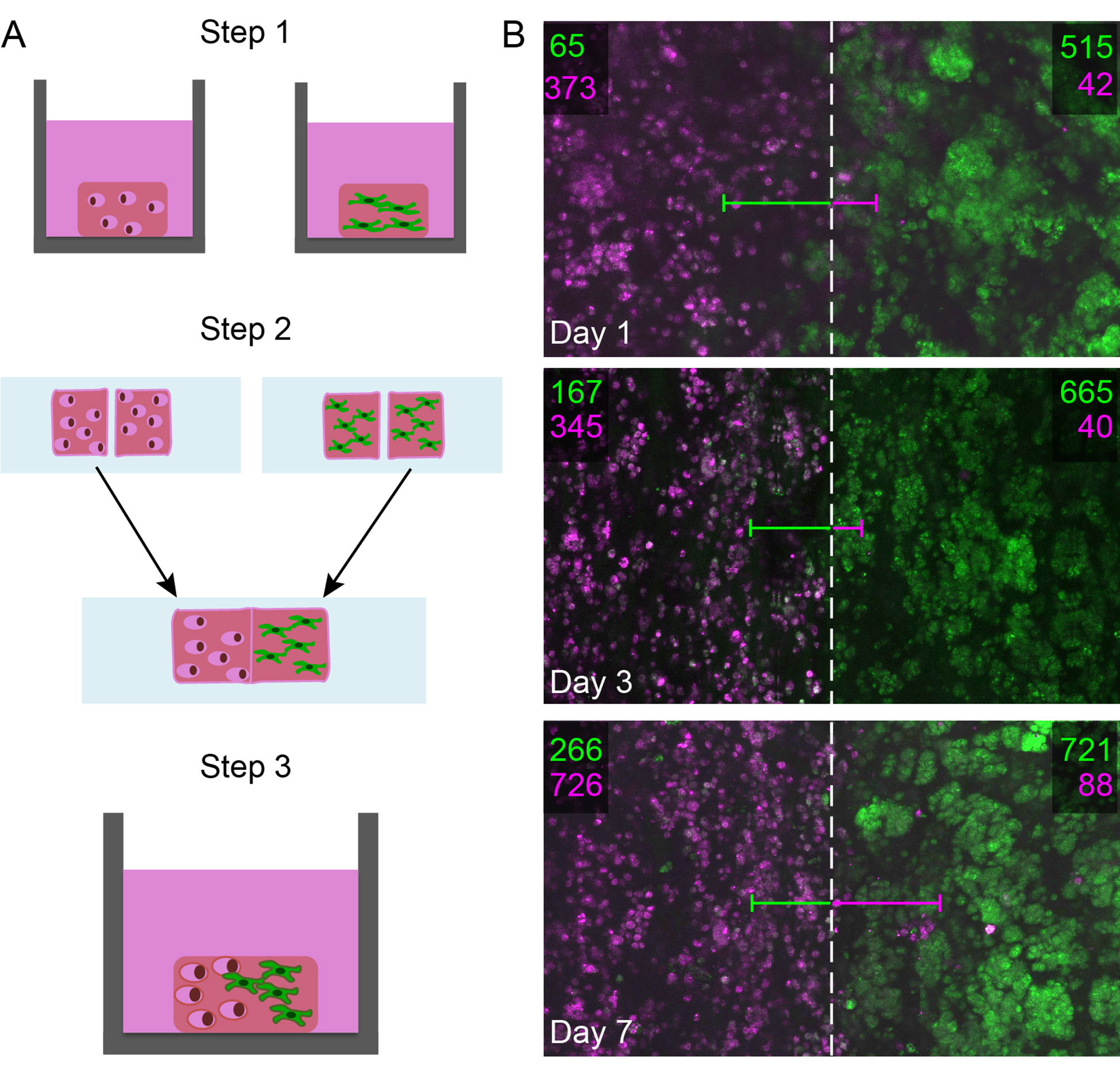
Figure 3.

A) Human pulmonary fibroblasts (CCL151, green) and human breast cancer cells (MDA MB 231, pink) were co-cultured in self-healing boronic acid-based hydrogels. B) Confocal z-stacks centered at the healed interface were taken over a large area of the healed hydrogel using a tile scan. The number of cells in a 1000 μm long region of interest on either side of the healing interface was quantified in three dimensions, where the number of fibroblasts is shown in green and the number of breast cancer cells is shown in pink for each side. The average distance each cell type had traveled across the interface into the opposite half is represented by each line (green line for fibroblasts and pink line for breast cancer cells). Projections of the confocal z-stack tile scan are shown here for a top down view of the 2000-μm long total region of interest (interface noted with a dashed lined).
