Fig. 1.
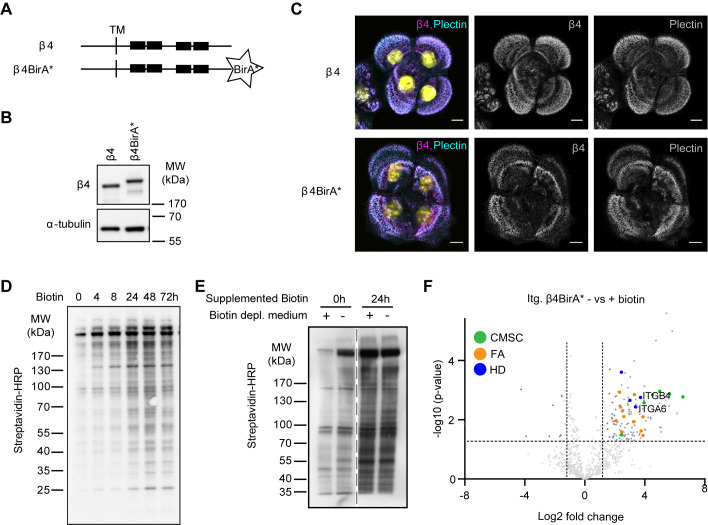
BioID method to identify β4 proximity interactors. (A) Schematic representation of wild-type β4 and the β4-BirA* fusion proteins. Black boxes indicate the FnIII domains. TM is transmembrane domain. (B) Western blot analysis of whole cell lysates from PA-JEB/β4 and PA-JEB/β4-BirA* keratinocytes probed with anti-β4 and anti-α-tubulin (loading control) antibodies. (C) Representative confocal microscopy images of PA-JEB keratinocytes expressing wild-type β4 and β4-BirA* stained for β4 and plectin. Scale bars: 10 μm. (D) Whole cell lysates from PA-JEB/β4-BirA* keratinocytes treated for the indicated time points with 50 μM biotin and analyzed by western blot with streptavidin-HRP. (E) Western blot analysis of biotinylated proteins from PA-JEB/β4-BirA* cells, cultured in regular medium or biotin-depleted medium for 20 h and subsequently treated with or without biotin for 24 h. Biotinylated proteins were detected by probing the membrane with Streptavidin-HRP. (F) Volcano plot showing enrichment (log2) and corresponding significance (P-value, log10) of biotinylated proteins in biotin-treated and -untreated PA-JEB/β4-BirA* keratinocytes (n=3).

