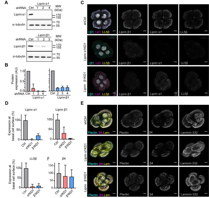
Fig. 4.
CMSCs are not required for the formation of HDs in keratinocytes. (A) Western blot analysis of stable shRNA-expressing PA-JEB/β4 cell lines [control (Ctrl) and three knockdowns (KDs)] probed with antibodies against liprin α1, liprin β1 and α-tubulin. (B) Quantification of liprin protein expression normalized to α-tubulin protein expression levels in knock down and control PA-JEB/β4 keratinocytes. Mean+s.d., n=2. (C) Triple immunofluorescence detection of liprin α1, liprin β1 and LL5β in liprin α1 and β1 knockdown and control keratinocytes. Scale bars: 10 μm. (D) Quantification of immunofluorescence staining of liprin α1, liprin β1 and LL5β in knockdown and control PA-JEB/β4 keratinocytes (n=20). (E) Triple immunofluorescence detection of β4, plectin and laminin-332 in liprin α1 and β1 knockdown and control PA-JEB/β4 keratinocytes. Scale bars: 10 μm. (F) Quantification of immunofluorescence staining of β4 shows no significant difference (Mann–Whitney test used) between liprin α1 and β1 knockdown and control PA-JEB/β4 keratinocytes (n=22).

