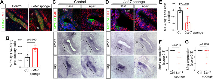
Fig. 3.
Disruption of let-7 miRNA function increases pro-sensory cell proliferation and delays HC formation in the developing chicken BP. Shown are adjacent sections of control BPs (A,C) and of corresponding let-7 sponge-treated BPs (A,D) 44 h after electroporation and EdU treatment at E4.5. Black bars indicate pro-sensory domains. (A) Mid-apical cross-sections of let-7 sponge-treated BP (A, right panel) and corresponding control BP (A, left panel) labeled for EdU (red) and the pro-sensory/sensory marker SOX2 (green). let-7 sponge treatment increased cell proliferation inside and outside the pro-sensory/sensory domain. (B) Quantification of A. Data are mean±s.d. (n=5 animals per group). (C,D) Adjacent cross-sections through the base and mid-apex of control (C) and let-7 sponge-treated (D) BPs were analyzed for MYO7A protein expression using immunostaining, and for Atoh1 and Lfng expression using RNA in situ hybridization. (E) Quantification of MYO7A+ HCs in control (C) and let-7 sponge-treated BPs (D). Individual data points represent average value per animal and treatment. Data are mean±s.d. (n=9 animals per group). (F,G) Quantification of Atoh1 (F) and Lfng (G) mRNA expression using a scoring system: 0, absent; 1, weak; 2, reduced; 3, normal expression. Four or five adjacent Atoh1- and Lfng-labeled cross-sections each representing base, mid and mid-apex were analyzed per animal and treatment. Data are mean±s.d. (n=8 animals per group). P-values were calculated using unpaired two-tailed Student's t-test (P≤0.05 is deemed significant). Ctrl, control; sac, sacculus; n, neural; a, abneural. Scale bars: 100 µm.

