Fig. 7.
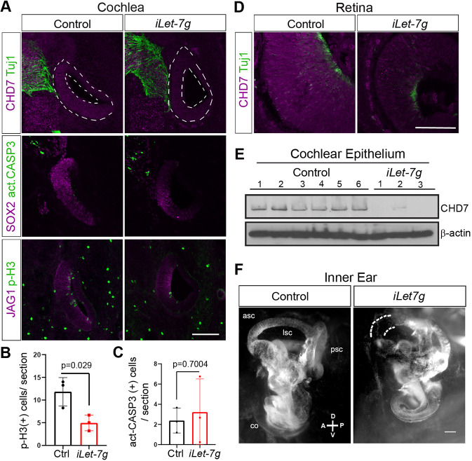
let-7g overexpression inhibits CHD7 expression in the developing murine retina and cochlea, and alters inner ear morphology. We administered dox to pregnant dams starting at stage E10.5 (A-E) or E11.5 (F) and harvested iLet-7g transgenic embryos and their single transgenic control littermates for further analysis at indicated stages. (A-C) let-7g overexpression reduces cochlear epithelial cell proliferation and cochlear pro-sensory CHD7 protein expression. (A) Confocal images of adjacent cochlear sections obtained from E13.5 iLet-7g embryos and control littermates. CHD7 (magenta) marks cochlear and neuronal progenitors; Tuj1 (green) marks neurons; SOX2 and JAG1 (magenta) mark the cochlear pro-sensory domain; act-CASP3 (green) labels apoptotic cells; pH3 (green) labels mitotic cells. Dashed white lines outline cochlear epithelia. (B,C) Quantification of the number of mitotic (pH3+) (B) and apoptotic (act-CASP3+) (C) cells in control (Ctrl, black bar) and let-7g overexpressing (iLet-7g, red bar) cochlear epithelia at stage E13.5. Data are expressed as mean±s.d. (n=3 animals per group, P-value calculated using unpaired two-tailed Student's t-test, P≤0.05 deemed significant). Scale bar: 100 µm. (D) Let-7g overexpression reduces retinal CHD7 protein expression. Confocal images of CHD7 and Tuj1 immunostained retinal sections from stage E13.5 iLet-7g embryos and control littermates. Scale bar: 100 µm. (E) let-7g overexpression reduces cochlear epithelial CHD7 protein expression. Western blot analysis of CHD7 protein expression in E15.5 control and let-7g overexpressing cochlear epithelia. β-Actin was used as loading control (n=6 animals for control and n=3 animals for iLet-7g group). (F) let-7g overexpression alters inner ear morphology. Paint-fills of E17.5 inner ears obtained from an iLet-7g transgenic embryo and a non-transgenic (control) littermate. The let-7g-overexpressing inner ear is smaller, has a shortened cochlea and malformed semicircular canals, with the anterior semicircular canal being truncated (dashed white lines) and the lateral semicircular canal being missing. asc, anterior semicircular canal; psc, posterior semicircular canal; lsc, lateral semicircular canal; co, cochlea. Scale bar: 200 µm.

