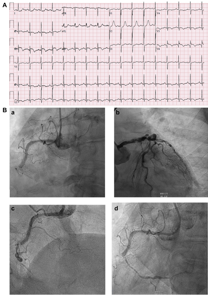
Figure 2.
Delayed Presentation Inferolateral Wall MI
(A) A 12-lead electrocardiogram showing inferoposterior ST-segment elevation myocardial infarction with posterolateral infarct pattern. (B) (a) RCA occlusion; (b) severe distal left main disease, proximal LAD disease, and occluded LCX; and (c and d) post wiring improved TIMI flow grade 3, revealing a severely calcified mid-RCA lesion. (C) Basal inferolateral pseudoaneurysm and ventricular septal rupture. LCX = left circumflex artery; TIMI = Thrombolysis In Myocardial Infarction; other abbreviations as in Figure 1.


