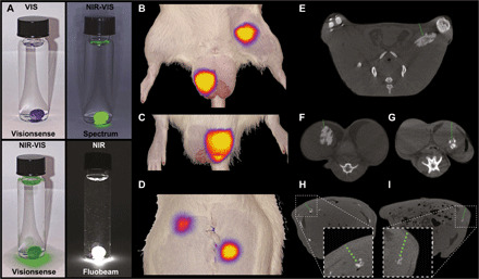
Fig. 4. Fluorescence imaging of XPVN-mark.

NIR fluorescence (NIR) and visual (VIS) light imaging of XPVN-mark (300 μl of XPVN in water) using commercially available NIR cameras: (i) Visionsense Iridium, (ii) Quest Spectrum, and (iii) Fluobeam, Fluoptics (A). XPVN-mark was injected into the thigh (100 μl) and right testicle (100 μl) (B) and left testicle (50 μl) (C) of a rat and NIR-VIS imaged (Visionsense Iridium). The abdominal cavity was surgically opened, and 10 μl of XPVN-mark was injected into the liver (top) and spleen (bottom) and NIR-VIS imaged (Visionsense Iridium) after surgically closing the abdominal wall (D). Corresponding axial-sliced CT images of the injected markers in thigh (E), testicles (F and G), liver (H), and spleen (I). The green dashed lines indicate the tissue depth (E: 5 mm, F: 2 mm, G: 6 mm, H: 5 mm, and I: 6 mm).
