
Fig. 6. Transactivation of Opn1mw in Rho+/− mice reduces apoptosis.
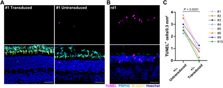
(A) Representative sections of the immunolabeled retina from Rho+/− mouse #1 injected with split dCas9-VPR and Opn1mw-specific sgRNAs showing a transduced (left) or untransduced (right) area of the same retina 1 year after injection. (B) Immunolabeling of the rd1 mouse retina on P13 served as a positive control. TUNEL staining (magenta, top) was used to visualize apoptosis, PRPH2 (cyan) was used as rod and cone outer segment marker (bottom). Scale bar, 30 μm. (C) Quantification of TUNEL+ cells in transduced versus untransduced areas of retinas from eight Rho+/− mice injected with split dCas9-VPR and Opn1mw-specific sgRNAs. A paired t test (two-tailed) was used for statistical analysis.