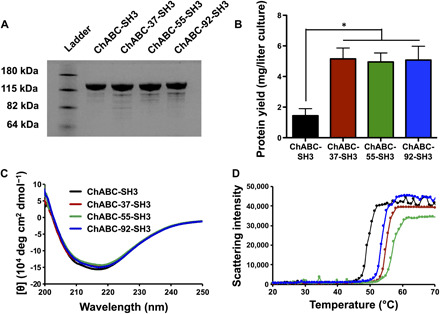
Fig. 3. ChABC-SH3 designs are highly expressed and more stable than wild type.

(A) Gel electrophoresis of 5 μg of ChABC-SH3 and mutated designs followed by Coomassie Brilliant Blue staining of protein bands. (B) ChABC-SH3 and mutant yield from large volume (2 liters) E. coli cultures (n = 3, means ± SD, *P < 0.05). (C) Circular dichroism spectra of ChABC-SH3 and designs from 200 to 250 nm at 25°C. (D) Protein aggregation curves between 20° and 70°C (1°C/min) measured by scattering intensity of the solution.
