Fig. 2A-B.

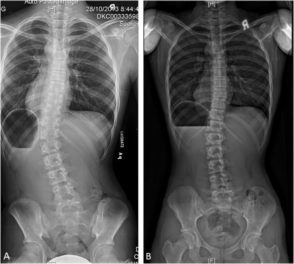
These radiographs are of a patient with improved curve magnitude, with (A) a pre-brace standing radiograph showing a T10 to L3 curve of 30° and (B) a post-brace standing radiograph showing curve regression of 15°.

These radiographs are of a patient with improved curve magnitude, with (A) a pre-brace standing radiograph showing a T10 to L3 curve of 30° and (B) a post-brace standing radiograph showing curve regression of 15°.