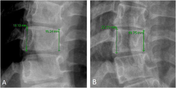
Fig. 4.

These radiographs are of a patient with curve progression and increased vertebral wedging as shown by (A) a pre-brace T10 apical ratio of 1:1 (convex height of 18 mm and concave height of 16 mm) and (B) a post-brace T10 apical ratio of 1:2 (convex height of 22 mm and concave height of 19 mm).
