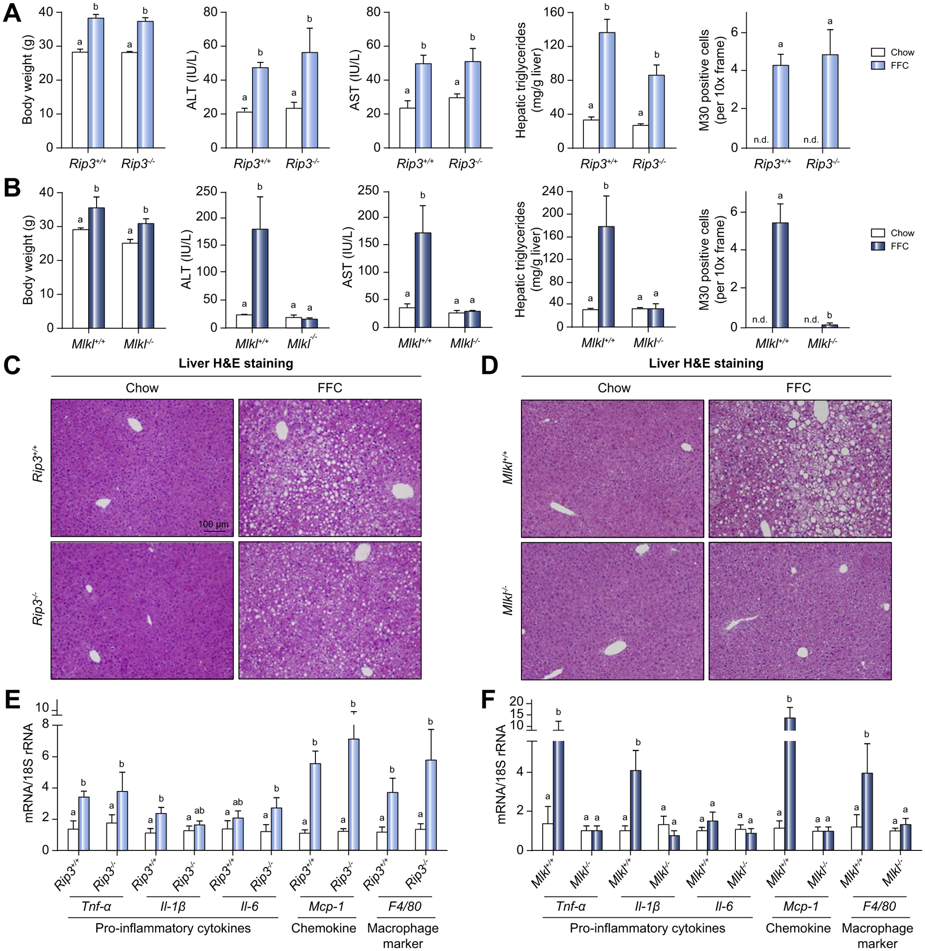
Fig. 1. Differential role of Rip3 and Mlkl deficiency on FFC diet-induced liver injury, steatosis, inflammatory response and hepatocyte apoptosis.

Rip3+/+, Rip3−/−, Mlkl+/+ and Mlkl−/− mice were allowed free access to FFC or chow diet for 12 weeks. Body weight, ALT and AST concentration in plasma, hepatic triglyceride content in whole liver homogenate and M30 positive cells (total number of cells per 10× frame) in formalin-fixed paraffin-embedded sections of liver from (A) Rip3+/+ and Rip3−/− and (B) Mlkl+/+ and Mlkl−/− littermates. Images of M30 are shown in Fig. S2. N.D.: M30-positive cells were not detectable in livers from chow-fed mice. H&E staining of livers from (C) Rip3+/+ and Rip3−/− and (D) Mlkl+/+ and Mlkl−/− mice on FFC or chow diet. Images were acquired at 10× magnification. Expression of mRNA for pro-inflammatory cytokines, chemokine and macrophage markers was detected in livers from (E) Rip3+/+ and Rip3−/− and (F) Mlkl+/+ and Mlkl−/− littermates using qRT-PCR and normalized to 18S rRNA. Values represent means ± SEM. Values with different superscripts are significantly different from each other, n = 3–6 per group. p <0.05, assessed by ANOVA. ALT, alanine aminotransferase; AST, aspartate aminotransferase; FFC, high-fat, high-fructose, high-cholesterol; qRT-PCR, quantitative reverse transcription PCR.
