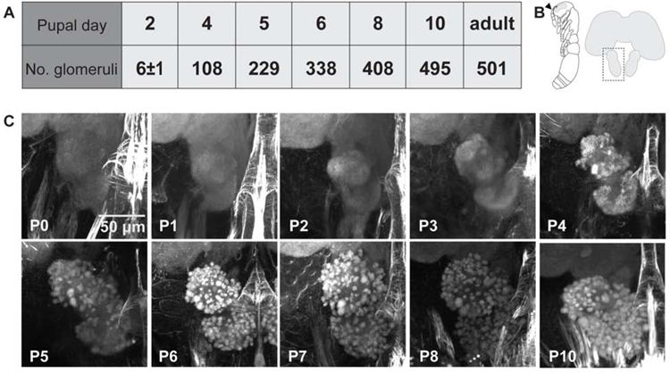
Figure 1: Antennal lobe development over the first 10 days of pupation.
(A) Manual counts of glomeruli from confocal stacks of pupal brains stained with phalloidin. Glomeruli were counted in the right antennal lobe from each brain. P2 n=6 brains with average 6±1 SD glomeruli; n=1 for all other pupal time-points; adult n=2 brains with 493 and 509 glomeruli (average 501; adult counts were previously published[13]). (B) Illustration of an ant pupa with the antenna marked by an arrowhead (left), and schematic of an ant brain with the left antennal lobe boxed to show the orientation of images in (C). (C) Representative projections of confocal z-stacks through the right antennal lobe of phalloidin stained pupal brains (see also Figure S1A).

