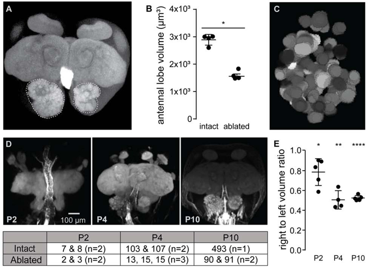
Figure 3: Early antenna ablation rapidly retards size and complexity of the developing antennal lobe.
(A) Maximum projection of a phalloidin-stained adult brain from an ant with the right antenna surgically removed at P0. Antennal lobes are outlined for context. (See also Figure S4.) (B) Antennal lobe volumes in adults were significantly lower on the side where antennae had been ablated at P0 compared to the AL with intact innervation (t-test for difference between average volumes). (C) Representative manual reconstruction of the right antennal lobe in (A) showed 91 distinct glomeruli. Reconstruction of a second brain (not shown) revealed 90 glomeruli (see also Figures S3–S6). (D) Representative images of developing pupa brains with the right antenna surgically removed at P0, along with glomerular counts at each time-point for each antennal lobe listed in table below. (E) Ratio of antennal lobe volumes measured from confocal z-stacks at each time-point showed volume of intact antennal lobes rapidly outpacing that of denervated lobes (t-test for difference from a volume ratio of one). *: p <0.05, **: p <0.01, ****: p <0.00001.

